A common cause of shoulder pain, irreparable rotator cuff tears are challenging to treat, and surgeons currently disagree on the most effective approach to treatment. Before a consensus can be reached on the optimal repair techniques for massive rotator cuff tears, more high-quality comparative studies are needed.
Toward that end, Christopher C. Schmidt, MD, is continually working to advance his field and improve the management of irreparable rotator cuff tears. Widely considered to be one of the best shoulder surgeons in the nation, Dr. Schmidt practices and performs extensive research at his biomechanical lab in Pittsburgh, Pennsylvania, where he has achieved many significant breakthroughs. Through these landmark efforts, Dr. Schmidt continues to improve patient care through evidence-based medicine, and his findings are regularly incorporated into guidelines used by his peers across the nation.
Pinpointing the Humeral Insertion Points for the Rotator Cuff Tendons
Recently, Dr. Schmidt participated in a research initiative designed to provide further insights into the geometry of the rotator cuff humeral footprints. This information is important for two main reasons: first, to provide surgeons with a better understanding of irreparable rotator cuff tears and rotator cuff function, and second, to help explain the improved outcomes experienced by patients who undergo superior capsule reconstruction.
The abstract of the study is reproduced below:
Morphology of the Rotator Cuff Humeral Footprints
Michael Smolinski1, Sean Delserro1, Patrick J. McMahon2, Mark C. Miller1, Patrick Smolinski1,3, Christopher Schmidt1,3
1 Department of Mechanical Engineering and Material Science, University of Pittsburgh, PA
2 Veterans Administration Pittsburgh Healthcare System, Pittsburgh, PA
3 Department of Orthopaedic Surgery, University of Pittsburgh, Pittsburgh, PA
Disclosures: None.
INTRODUCTION: Patient reported outcomes and active range of motion have been shown to improve following reconstruction of the superior capsule and rotator cuff for treatment of irreparable tears [1]. However, no studies have used advanced measurement techniques of anatomic dissections to describe the insertions of the rotator cuff. The goal of this study was to locate and precisely measure the humeral insertions of the rotator cuff tendons.
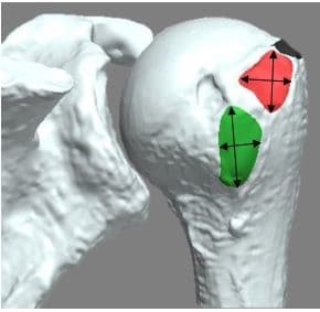
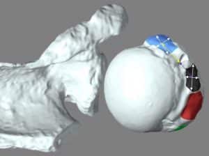
METHODS: With approval from the institutional review board, nine (n=9) fresh-frozen cadaveric specimens (average age 67.3 ± 2.6 yrs) were used in this study. Each rotator cuff tendon, the supraspinatus (SS), infraspinatus (IS), teres minor (TM), and subscapularis (SubS), was dissected individually from the superior capsule from origin to insertion. Following the transection of each tendon, the outline of the footprint was carefully marked with ink. A laser scanning system (Faro, Inc.) was used to make three-dimensional models of each specimen [2]. Using a probe, points along outline of each footprint were taken and incorporated into the models. The surface area for each footprint was calculated and measurements along the surface of the major (largest) dimension, the perpendicular dimension, and distance from the centroid to the apex of the bicipital groove were taken (Figures 1,2).
RESULTS: Measurements of the surface area, major and perpendicular dimensions, and distance of the centroid to the bicipital groove for the tendon insertion sites given in Table 1.
DISCUSSION: When compared to the supraspinatus, the teres minor, infraspinatus, and subscapularis have larger insertion areas on the proximal humerus.
SIGNIFICANCE/CLINICAL RELEVANCE: Information on the geometry of the rotator cuff insertions can provide surgeons with a better understanding of function and injury and offer an explanation for the improved outcomes of patients who undergo superior capsule reconstruction.
REFERENCES: [1] Mihata, et al., Arthroscopy, 2013 [2] Fujimaki, et al., AJSM, 2016
IMAGES AND TABLES:
Figure 1: Posterior view showing the measured dimensions of infraspinatus (IS-red) and teres minor (TM-green)

Figure 2: Superior view showing the measured dimensions of supraspinatus (SS-black) and subscapularis (SubS-blue), and the distance to the apex of bicipital groove.

Table 1: Measurements of insertion site dimensions, position and area (Mean±S.D).

If you have questions about this study or about irreparable massive rotator cuff tears in general, call (877) 471-0935 to schedule a consultation with Dr. Schmidt at one of his office locations in the greater Pittsburgh, PA, area.


